Content
Peritonitis in cattle is characterized by stagnation of bile due to blockage or compression of the bile duct. The disease often develops in cows after suffering pathologies of other organs, as well as some infectious diseases. Peritonitis has clear clinical signs, various forms and stages of manifestation. Diagnosis is made based on symptoms and laboratory tests.
What is peritonitis
Peritonitis is diffuse or limited inflammation of the parietal and visceral layers of the peritoneum, which may be accompanied by active exudation. It occurs in many representatives of the animal world, but more often birds, horses and cattle suffer from it. According to its etiology, the disease can be infectious and non-infectious, that is, aseptic, as well as invasive. According to localization, it can be diffuse, limited, and according to its course - acute or occurring in a chronic form. Peritonitis is also distinguished by the nature of the exudate released. It can be serous, hemorrhagic and purulent. Sometimes the disease has mixed forms.
The peritoneum is the serous covering of the walls and organs of the abdominal cavity. Moving from the walls to the internal organs, it forms folds and ligaments that limit the space. As a result, pockets and sinuses are formed.Essentially, the peritoneum is a kind of membrane that performs a number of functions, mainly as a barrier. The abdominal cavity is bounded above by the diaphragm, below by the pelvic diaphragm and pelvic bones, behind by the spine, lumbar muscles, and on the sides by the oblique and transverse muscles.
Causes of peritonitis in cattle
The acute course of the disease in cattle develops after trauma to the gastrointestinal tract (perforation by foreign objects, rupture, perforated ulcer), uterus, urinary and gall bladder. Chronic peritonitis, as a rule, persists after an acute process or occurs immediately with tuberculosis or streptotrichosis. Sometimes it occurs in a limited area, for example, as a result of an adhesive process.
Peritonitis of an infectious-inflammatory nature occurs after appendicitis, cholecystitis, intestinal obstruction, vascular thromboembolism, and various tumors. Traumatic peritonitis occurs with open and closed wounds of the abdominal organs, with or without damage to the internal organs. Bacterial (microbial) peritonitis can be nonspecific, caused by the intestinal microflora, or specific, which is caused by the penetration of pathogenic microorganisms from the outside. Aseptic peritonitis occurs after exposure of the peritoneum to non-infectious toxic substances (blood, urine, gastric juice).
In addition, the disease can be caused by:
- perforation;
- surgical intervention on the peritoneal organs with an infectious complication;
- use of certain medications;
- penetrating abdominal wound;
- biopsy.
Thus, the disease occurs as a result of pathogenic microorganisms entering the peritoneal area.
Symptoms of peritonitis in cattle
The following manifestations of the disease are characteristic of cattle with peritonitis:
- increased body temperature;
- lack or decreased appetite;
- increased heart rate and breathing;
- on palpation, pain in the abdominal wall;
- gases in the intestines, constipation;
- dark-colored stool;
- vomit;
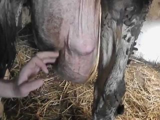
- sagging abdomen due to fluid accumulation;
- slowing down or stopping the work of the rumen;
- yellowness of the mucous membranes;
- hypotension of the preventricles;
- agalaxia in dairy cows;
- depressed state.
With putrefactive peritonitis in cattle, the symptoms are more pronounced and develop faster.
Laboratory blood tests show leukocytosis and neutrophilia. Urine is dense and high in protein. On rectal examination, the veterinarian detects focal tenderness. In addition, gases in the intestines are noted in the upper part of the abdominal cavity, and exudate in the lower part.
Chronic peritonitis of diffuse form occurs with less pronounced symptoms. The cow loses weight, sometimes has a fever, and attacks of colic occur. Exudate accumulates in the peritoneal cavity.
With a limited chronic disease in cattle, the function of nearby organs is impaired. Gradually, cows lose fat.
Peritonitis in cattle is characterized by a protracted course. Acute and diffuse forms of the disease are sometimes fatal within a few hours of the onset of symptoms. The chronic form can last for years. The prognosis in most cases is unfavorable.
Diagnostics
The diagnosis of peritonitis in cattle is made based on the clinical manifestations of the disease, laboratory blood tests, and rectal examination. In doubtful cases, fluoroscopy, laparotomy, and a puncture from the peritoneal cavity are performed.A veterinarian should rule out fasciliosis, ascites, obstruction, and diaphragmatic hernia in cattle.
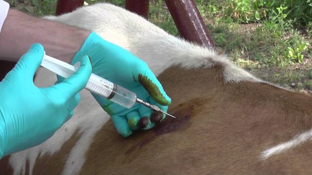
A puncture from cattle is taken from the right side near the ninth rib, a few centimeters above or below the mammary vein. To do this, use a ten-centimeter needle with a diameter of 1.5 mm.
Fluoroscopy can detect the presence of exudate in the abdominal cavity and air.
Using laparoscopy, the presence of adhesions, neoplasms, and metastases is determined.
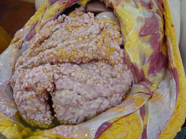
When opening an animal that has died from peritonitis, a hyperemic peritoneum with pinpoint hemorrhages is discovered. If the disease began not so long ago, then serous exudate is present; with further development of peritonitis, fibrin will be detected in the effusion. The internal organs in the abdominal cavity are glued together by a protein-fibrous mass. Hemorrhagic peritonitis is found in some infections and in mixed forms of the disease. Purulent, purulent exudate is formed when the intestines and proventriculus rupture. In case of chronic cattle peritonitis, after injury, connective tissue adhesions of the peritoneal layers with the membranes of the internal organs are formed.
Treatment of peritonitis in cattle
First of all, the animal is prescribed a starvation diet, cold wraps are applied to the abdomen, and complete rest is provided.
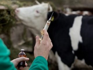
Drug therapy will require antibiotics and sulfonamides. To reduce vascular permeability, reduce fluid secretion, and relieve symptoms of intoxication, a solution of calcium chloride, glucose, and ascorbic acid is prescribed intravenously. To relieve pain, a blockade is performed using the Mosin method. For constipation, you can give an enema.
The second stage of therapy is aimed at accelerating the resorption of exudate. For this purpose, physiotherapy and diuretics are prescribed. In severe cases, puncture suction is performed.
If a wound surface or a scar serves as a gateway for infection to enter the abdominal cavity of cattle, then it is cut, cleaned, tamponed with sterile gauze and disinfected.
Preventive actions
Prevention is aimed at preventing diseases of the abdominal organs, which can contribute to the development of secondary peritonitis in cattle. It is recommended to follow the basic standards of care and maintenance of livestock and to prevent foreign bodies from getting into the feed. To do this you need to use:
- magnetic separator for cleaning feed;
- veterinary indicator that determines the position of an object in the cow’s body;
- a magnetic probe that can be used to remove foreign bodies;
- cobalt ring that prevents stomach injuries in cattle.
Conclusion
Peritonitis in cattle is a serious disease of the peritoneum that occurs as a complication after pathologies of nearby organs. The causes of peritonitis are varied. The clinical picture of the disease depends on the course and form of the disease. A conservative treatment method can help if the diagnosis is correct and therapy is started on time. Otherwise, peritonitis in cattle most often ends in death.

