Content
Due to improper maintenance and inadequate diet, breeding farm animals are often overtaken by various non-contagious diseases associated with impaired metabolism or general muscle weakness. One of these diseases, myopathy or white muscle disease of calves, is very common in cattle. Calves are not the only ones suffering from this disease. Myopathy was recorded not only in all types of livestock, but even in poultry.
What is white muscle disease
Myopathy is a non-contagious disease of young animals. Most common in countries with developed cattle breeding:
- Australia;
- USA;
- New Zealand.
Beef from these countries is exported all over the world, but inferior feed is used to reduce the cost of production. Such nutrition promotes the growth of muscle mass, but does not provide animals with all the necessary elements.
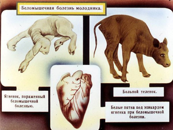
White muscle disease is characterized by deep structural and functional disorders of the myocardium and skeletal muscles. As the disease progresses, the tissues become discolored.
Myopathy occurs in areas with sandy, peat and podzolic soils poor in microelements.
Causes
The etiology of myopathy has not yet been studied, although it has been known about it for more than 100 years. Main version: lack of micro- and macroelements, as well as vitamins in animal feed. But it has not yet been determined which element should be added to the feed in order to avoid myopathy.
The main version of the occurrence of white muscle disease in young animals is a lack of selenium, vitamin A and protein in the uterine feed. The baby did not receive these substances in the womb and does not receive them after birth. This situation can arise even on free grazing if there is a lot of sulfur in the soil. This element interferes with the absorption of selenium. If sulfur dissolves in the soil after rains and plants absorb it, animals may develop a “natural” selenium deficiency.
The second version: myopathy occurs when there is a lack of a whole complex of substances at once:
- Selena;
- iodine;
- cobalt;
- manganese;
- copper;
- vitamins A, B, E;
- amino acids methionine and cysteine.
The leading elements in this complex are selenium and vitamin E.
Course of the disease
The insidiousness of white muscle disease is that its initial stage is invisible. This is the moment when the calf can still be cured. Once symptoms become apparent, treatment is often no longer useful. Depending on the form, the course of the disease may take more or less time, but the development is always increasing.
Symptoms of white muscle disease in calves
In the initial period, there are almost no external signs of white muscle disease, except for rapid pulse and arrhythmia. But few cattle owners measure the calf’s pulse every day. Then the animal begins to quickly get tired and move little.This is sometimes also attributed to a calm character.
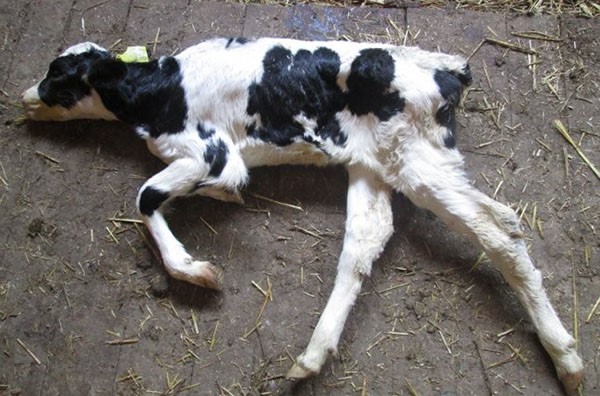
Myopathy is noticed when calves stop standing up and prefer to lie down all the time. By this time, their reflexes and pain sensitivity are noticeably reduced. The previously poor appetite disappears completely. At the same time, salivation and diarrhea begin. Body temperature is still normal, provided there is no bronchopneumonia as a complication. In this case, the temperature rises to 40-41 °C.
At the last stage of white muscle disease, the calf's pulse becomes weak to threadlike, and increases to 180-200 beats per minute. A clearly defined arrhythmia is observed. Breathing is shallow with a frequency of 40-60 breaths per minute. Exhaustion is progressing. A blood test shows the presence of vitamin deficiencies A, E, D and hypochromic anemia. The urine of a calf with myopathy is acidic with a large amount of protein and myochrome pigment.
The symptoms of various forms of myopathy are not fundamentally different from each other. Only their expression differs.
Acute form
The acute form is observed in newborn calves. It is distinguished by pronounced symptoms. The duration of white muscle disease in the acute form is about a week. If action is not taken immediately, the calf will die.
In the acute form, signs of white muscle disease appear very quickly:
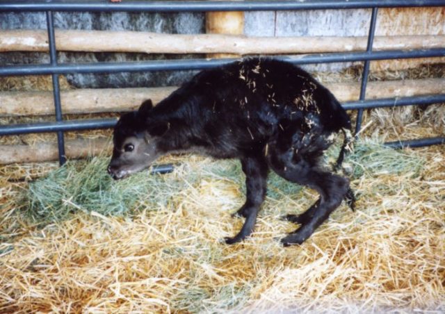
- the calf tries to lie down;
- muscle tremors occur;
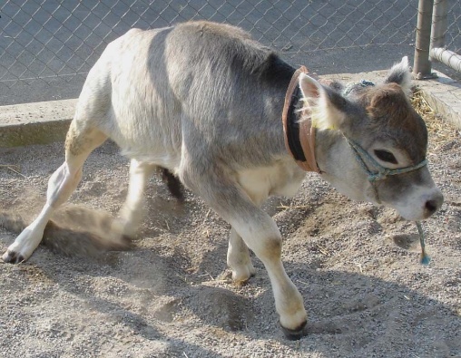
- gait is disturbed;
- paralysis of the limbs develops;
- breathing difficult, frequent;
- serous discharge from the nose and eyes.
The work of the gastrointestinal tract also begins to stop. Due to the stoppage, food decomposes in the intestines with the release of gases. External signs of stoppage include swollen intestines and foul-smelling feces.
Subacute forms
The subacute form differs only in more “smoothed out” symptoms and a longer course of the disease: 2-4 weeks. The owner is more likely to notice something is wrong and have time to take action. Due to this, deaths in the subacute form of myopathy account for 60-70% of the total number of sick calves.
Chronic form
The chronic form of myopathy occurs in calves older than 3 months. This form develops gradually due to an unbalanced diet, in which the necessary elements are present, but in small quantities. Due to mild symptoms, the disease can progress to irreversible changes in the muscle structure. In the chronic form, animals are exhausted, inactive and retarded in development. Sometimes calves' hind legs fail.
Diagnostics
The primary lifetime diagnosis is always presumptive. It is placed on the basis of the enzootic development of the disease and its stationarity. If white muscle disease has always occurred in a given area, then in this case it is also with a high degree of probability. Also auxiliary signs are the clinical picture and myochrome in the urine.
Modern diagnostic methods also allow intravital fluoroscopy and electrocardiography. But such studies are too expensive for most farmers, and not all veterinarians can correctly read the results. It's easier to slaughter one or two calves and do a necropsy.
An accurate diagnosis is made after an autopsy based on characteristic pathological changes:
- softening of the brain;
- swelling of the fiber;
- skeletal muscle dystrophy;
- the presence of discolored spots on the myocardium;
- enlarged lungs and heart.
Myopathy in calves is differentiated from other non-communicable diseases:
- rickets;
- malnutrition;
- dyspepsia.
Case histories here are similar to white muscle disease in calves and originate in an unbalanced diet and improper feeding. But there are also differences.
Rickets has other characteristic manifestations that affect the musculoskeletal system:
- curvature of bones;
- joint deformation;
- spinal deformity;
- osteomalacia of the chest.
Rickets is similar to myopathy due to the exhaustion of the calf and disturbances in gait.
Signs of malnutrition are similar to white muscle disease in the area of general underdevelopment and weakness of skeletal muscles. But it does not cause irreversible changes in the heart muscle.
With dyspepsia, the calf's stomach swells, diarrhea, dehydration and general intoxication may occur. Muscle dystrophy is not observed.
Treatment of white muscle disease in calves
If symptoms are recognized early and treatment for white muscle disease in calves is started at an early stage of development, the animal will recover. But if signs of heart block and myocardial dystrophy are already obvious, it is useless to treat the calf.
Sick calves are placed in a dry room on soft bedding and placed on a milk diet. Also included in the diet:
- quality hay;
- grass;
- bran;
- carrot;
- oatmeal;
- pine infusion;
- vitamins A, C and D.
But such a diet, in addition to the pine infusion, should be normal when feeding a calf. Therefore, in the treatment of white muscle disease, this is an important, but not the only complex.
In addition to the diet, additional microelements are used to treat myopathy:
- subcutaneously 0.1% selenite solution at a dose of 0.1-0.2 ml/kg body weight;
- cobalt chloride 15-20 mg;
- copper sulfate 30-50 mg;
- manganese chloride 8-10 mg;
- vitamin E 400-500 mg daily for 5-7 days;
- methionine and cysteine 0.1-0.2 g for 3-4 days in a row.
Instead of giving it with food, vitamin E is sometimes administered as injections of 200-400 mg for 3 days in a row and another 4 days for 100-200 mg.
In addition to microelements for myopathy, heart medications are also given:
- cordiamine;
- camphor oil;
- subcutaneously tincture of lily of the valley.
If complications occur, sulfonamides and antibiotics are prescribed.
Forecast
In the early stages of the disease, the prognosis is favorable, although the calf will lag behind in development and body weight gain. It is not advisable to leave such animals. They are raised and slaughtered for meat. If the disease is advanced, it is easier and cheaper to kill it right away. Such a calf will not grow, and in especially severe cases it will die due to irreversible changes in the myocardial tissue.
Prevention measures
The basis for the prevention of white muscle disease in calves is proper maintenance and feeding of animals. The diet of pregnant cows is tailored to local conditions and soil composition. Feeds must be balanced. Their composition must contain in sufficient quantities:
- proteins;
- sugar;
- vitamins;
- micro- and macroelements.
To ensure the desired composition, the necessary additives are added to the feed mixture. For this reason, feed must be periodically sent for chemical composition analysis. With systematic analyses, the composition of the feed can be quickly adjusted.
In disadvantaged areas, queens and offspring are treated with selenite preparations. Cattle are injected subcutaneously with 30-40 mg of sodium selenite solution 0.1%. Injections begin in the second half of pregnancy and are repeated every 30-40 days. Stop injecting selenite 2-3 weeks before calving. Calves are injected with 8-15 ml every 20-30 days.
Sometimes it is recommended to use tocopherol together with selenite.In addition, once a day they give other missing elements (adults and calves, respectively):
- copper sulfate 250 mg and 30 mg;
- cobalt chloride 30-40 mg and 10 mg;
- manganese chloride 50 and 5 mg;
- zinc 240-340 mg and 40-100 mg for calves up to 6 months;
- iodine 4-7 mg and 0.5-4 mg for calves up to 3 months.
The addition of elements is carried out only after a chemical analysis of the feed, since an excess is no less harmful than a deficiency.
Conclusion
White muscle disease in calves in the final stages is incurable. The easiest way to maintain livestock is to monitor the content and balance of the diet.
