Content
Actinomycosis in cattle is a disease known since the 1970s. The causative agent of the pathology was identified by the Italian scientist Rivolt. This discovery was later confirmed by German researchers. In the modern world, actinomycosis is spreading more and more, affecting a huge number of cattle. Read more about the symptoms, methods of diagnosis and treatment of the disease below.
What is actinomycosis in cattle?
Among cattle diseases, actinomycosis occupies a leading position. This disease has been known since ancient times. Scientists examined the jaws of a rhinoceros that lived in the Tertiary period. They found changes characteristic of actinomycosis.
The main target for infection is cattle. Sometimes pigs get sick, and most rarely other animals. Most often, the disease affects the following areas of the cow’s body:
- lower jaw;
- gum;
- sky;
- space between jaws;
- pharynx;
- The lymph nodes;
- salivary glands.
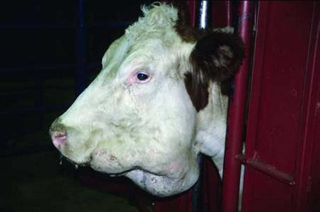
Separately, damage to the udder and tongue is distinguished. In the photo, actinomycosis of cattle looks like this.
Causes of cow disease actinomycosis
The causative agent of actinomycosis is the fungus Actinomyces bovis. In atypical cases, other types of fungus are isolated. In the exudate (inflammatory fluid), the pathogen is isolated in the form of small brown grains, which are also called drusen. They are gray or yellow in color.
When examining smears of sick cows under a microscope, the fungus looks like threads tangled together. Moreover, their diameter is uneven: there is a thickening at the periphery and a thin area in the middle.
But the fungus is not the only causative agent of actinomycosis. Sometimes when examining pus, bacteria are isolated:
- Pseudomonas aeruginosa;
- protea;
- staphylococci or streptococci.
Some researchers argue that actinomycosis is caused by an association of fungi and bacterial flora.
Actinomyces bovis actively develops under aerobic and anaerobic conditions. This means that the fungus does not care whether there is access to oxygen. When heated to 75°C, the causative agent of actinomycosis is destroyed within 5 minutes, and a formaldehyde solution kills it in 3 minutes. In the environment, actinomycetes survive for 2 years and are resistant to low temperatures.
The causative agent of actinomycosis enters the cow's body through damage to the skin, wounds in the mouth, and teats in the udder. The infection can enter through the respiratory tract and rectum. Sometimes cows become infected endogenously. Actinomycetes, which are present in the intestines and oral cavity of healthy cattle, are suddenly activated and cause an inflammatory process.
In the medical history of bovine actinomycosis, in most cases there is a history of trauma, which served as a gateway to infection. The source of actinomycosis infection can be feed, water, and other objects that cows interact with that are contaminated with pathogenic fungi.
From the gates of infection, the pathogen spreads through the connective tissue and subcutaneous fat. Therefore, actinomycosis is most often local in nature. Sometimes it spreads through the blood throughout the body.
Symptoms
Clinical manifestations of cow actinomycosis depend on the localization of the pathological process, the state of the animal’s immune system, and the aggressiveness of the pathogen. But all types of cow disease have several common features. Any form of actinomycosis is chronic. The disease begins with an incubation period. It represents a period of time when the pathogen is already active in the cow’s body, but clinical manifestations have not yet been observed.
Another common symptom is the formation of actinomyoma in cows. This is a large formation, which is essentially a benign tumor. It grows slowly, does not hurt, and has a dense consistency.

When the head is affected, cows develop dense nodes that grow both outward and inward, into the throat. Soon fistulas form on the actinomyomas. Through them, yellowish pus is released, which contains grains. These inclusions are drusen of the fungus. After a certain time, small areas of skin begin to die, so impurities of rejected tissue appear in the pus. The color of the discharge becomes reddish. The fistula opens up and then heals.
As the tumor grows in the throat, the cow begins to breathe with difficulty and has difficulty swallowing. As a result, due to a violation of the act of swallowing, the animal loses weight. Despite the abundant discharge of pus, the temperature most often remains normal. The increase is typical only for generalized actinomycosis.
When the jaws or the space between them are affected, the shape of the cattle's head changes.The jaw of cows increases several times. Sometimes the inflammation spreads to the surrounding tissues, causing fistulas (holes) to form in the palate and gums. A purulent mass flows out of them.
Actinomycosis of the cattle udder is characterized by a predominant lesion of the posterior lobes. It manifests itself as massive necrosis of the skin. First, dense ridges with a purulent cavity in the center form on the udder. Then, in their place, fistulas develop, from which a yellowish secretion flows.
Actinomycosis of the tongue is characterized by widespread or limited inflammation of this organ. People call it “wooden tongue.” Cows most often develop an ulcer on the back of the organ. The ulcer has a gray-white bottom with ridges along the edges.
Diagnostics
Treatment of actinomycosis in cows requires making a correct diagnosis. Most often it is not in doubt. A professional veterinarian may suspect actinomycosis based on clinical manifestations. But in any case, laboratory confirmation is necessary to select effective therapy.
Additional diagnosis consists of examining the pathological secretion under a microscope. For this purpose, pus, granulomatous tissue, and oropharyngeal smears are taken. Diagnosis of actinomycosis is carried out as follows:
- A secretion or part of a tubercle suspected of pathology is taken.
- Rinse them under water.
- Treat with an aqueous alkaline solution.
- Place on a glass slide.
- Fix with a 50% glycerin solution.
- Cover the top with a glass slide.
Only after all the preparatory stages have been completed can you be confident in the quality of the research. But the decisive factor in diagnosing actinomycosis is inoculating the pathological secretion on nutrient media. However, bacteriological research is difficult.
Determining the level of antibodies to the pathogen is not widespread in veterinary medicine, although it is widely used in diagnosing diseases in humans. Thus, the microscopy method is most often used.
When making a diagnosis, actinomycosis should be differentiated from other diseases of cows:
- actinobacillosis;
- streptotrichosis;
- foot and mouth disease;
- epizootic lymphangitis;
- tuberculosis of the lymph nodes.
Actinomycosis and actinobacillosis are most similar. But in the first case, the bone is most often damaged, in the second - the soft tissue of cows. Excellent pathogens under microscopic examination. The causative agent of actinomycosis has the form of long filaments, actinobacillosis - rods.
Tuberculosis of the lymph nodes differs from actinomycosis in that the first case is not characterized by the formation of an abscess. Cows infected with Mycobacterium tuberculosis react during tuberculinization.
How to treat actinomycosis in cows
The main goal of disease therapy is elimination of the pathogen. This implies the complete elimination of the fungus from the body of cattle.
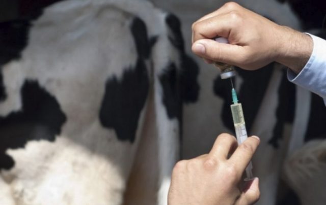
In the initial stage of the disease, iodine compounds are used. They are given to cows in the oral cavity and in the form of parenteral injections. Solutions of iodine and calcium iodide are infused intravenously. They are mixed with distilled water or physiological sodium chloride solution. For 1 ml of iodine take 2 ml of potassium iodide and 500 ml of water. But with this treatment, relapses of actinomycosis are possible.
To completely cure a cow, they turn to antibiotic therapy. The course of treatment is from 4 to 6 days. The duration depends on the severity of the disease, the state of the cow’s immunological resistance, and the resistance of the pathogen. The most commonly used drug is Oxytetracycline. When treating actinomycosis in adult cattle, the dose of the drug per application is 400,000 units; calves are given 200,000 units.
Actinomyomas are locally injected with Polymyxin. It also belongs to the group of antibiotics. 900 units are dissolved in 20 ml of novocaine. The latter is used for pain relief. The procedure is carried out once every 10 days.
The course of treatment should be strictly as determined by the specialist. As a rule, therapy is continued for several more days after the complete cessation of clinical manifestations. This is the only way to finally get rid of the pathogen.
When treating bovine actinomycosis with antibiotics, potassium iodide is used as a concomitant therapy. The dose for 1 administration is 100 ml of a 10% solution. Ultrasound irradiation is becoming increasingly widespread.
For localized forms of the disease, surgical removal of the tumor is considered the most effective. The actinonomicoma is excised completely along with the capsule. To increase the effectiveness of therapy, the cow is first given a course of antibiotics. Moreover, medications are administered both locally into the formation and intravenously. Below is a video of surgical treatment of bovine actinomycosis.
The room in which the sick cow was located must be disinfected. To do this, use a 3% alkaline solution or freshly slaked lime.
Inexpensive but effective drugs for the treatment of actinomycosis in cattle
Drugs for etiotropic therapy (aimed at eliminating the pathogen) are in most cases inexpensive. At the same time, they are highly efficient. To treat actinomycosis in cows, the following antibiotics can be used:
- "Penicillin";
- "Benzylpenicillin";
- "Okitetracycline";
- "Erythromycin";
- Metronidazole (effective for anaerobic infections).
Drugs from other groups are used together with antibiotics. Among antiseptics, the drug “Monoclavit-1” is highly effective. This product contains iodine. It is effective against both gram-positive and gram-negative bacteria. Its mechanism of action is the formation of a film on the surface of the wound, which protects it from environmental pollutants. External treatment is carried out once a day.
Zinaprim is another inexpensive but effective drug for the treatment of actinomycosis in cows. It is sold in powder form. The medicine is given to cows orally at the rate of 1 g per 10 kg of body weight. The course of treatment is from 3 to 5 days. "Zinaprim" also acts on gram-positive and gram-negative microorganisms. The medicine should not be given to cows with hypersensitivity to sulfamethazine, the active ingredient of the medicine.
The dietary supplement “Polyfit-propolis” cannot be ignored. The course of treatment with the drug is long. It ranges from 16 to 21 days. Therefore, it is used in combination with other medications.
Forecast
The prognosis of actinomycosis depends on the form of the disease, the severity of its course, the timeliness and adequacy of treatment of the cow. The earlier antibiotic therapy is started, the greater the likelihood of complete recovery of cattle without relapse.With localized forms, the prognosis is favorable. It worsens with generalized varieties of the disease or when joints are involved in the process.
Some treated cows develop reinfection. This is often due to inadequate antibiotic therapy. Livestock producers stop treatment immediately after symptoms disappear. As noted earlier, this is fundamentally false.
Preventive measures
Actinomycosis, like many other diseases of animals and humans, is easier to prevent than to cure. Therefore, it is very important to prevent this disease on farms. To reduce the risk of cows becoming infected, you must follow these rules:
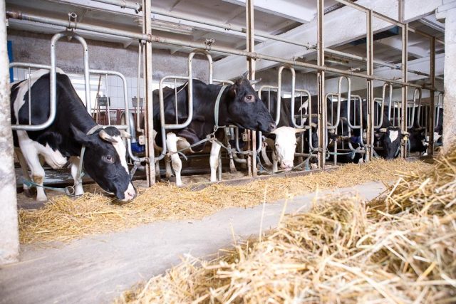
- Regularly disinfect cattle stalls. An effective treatment agent is freshly slaked lime.
- Do not graze cows in damp places or lowlands if the farm is located in an area unfavorable for the fungus.
- Prepare roughage before consumption by cows. To do this, pour boiling water over them and leave for 5-10 minutes. You can also add salt (10-15 g per 10 liters of water).
- Calcinate the straw before feeding the cow.
- Cattle that have already become infected with actinomycosis must be urgently isolated.
- Cows that have recovered from the disease must be under constant observation, as a relapse of the disease is possible.
Conclusion
Actinomycosis in cattle is a disease that requires the earliest possible diagnosis and treatment. With timely initiation of therapy, complete recovery of cows can be achieved. The main thing is not to self-medicate, but to seek help from a veterinarian.Only a specialist can determine the exact dosage and duration of the course.