Content
The most dangerous parasites of farm animals are tapeworms or tapeworms. They are dangerous not because they cause economic damage to livestock. Infected animals practically do not suffer from these types of worms. Humans suffer from them, as the ultimate host of the parasite. The larvae of one type of tapeworm cause fynosis in cattle and subsequent infection of humans with a long-lived worm up to 10 m long and a lifespan of 10 years. But using bull tapeworm is good for losing weight. You can eat anything and as much as you want. But this, of course, is sarcasm.
What is bovine cysticercosis?
A more correct name for bovine fynosis is cysticercosis. But Finnoz is easier to pronounce and remember.
The “ancestors” of cysticercosis are tapeworms of various species from the genus Tenia, also known as Cystodes. These parasites are most common in relatively warm regions:
- Africa;
- Philippines;
- Latin America;
- Eastern Europe.
But you can also meet them in Russia. Especially taking into account the widespread import of elite cattle breeds from Western countries to the Russian Federation.
Cattle are infected not by the helminths themselves, but by their larvae, which even have their own Latin name: personal for each species. Therefore, in essence, bovine cysticercosis is the infection of livestock with bovine tapeworm larvae.
Cattle may also contain larvae of other types of tapeworms, but their localization differs from the location of bovine cysticerci.
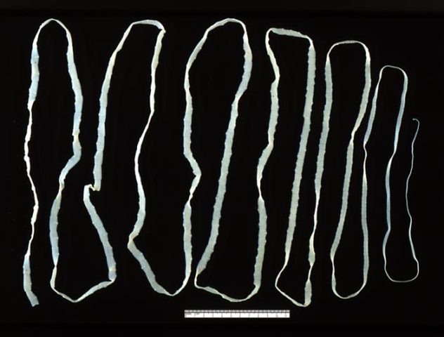
This is not a ribbon, but the “culprit” of cattle finosis - a bovine tapeworm, which is 10 m long. The head is on the right
Life cycle of tapeworm and infection of cattle with finnosis
The adult parasite can only live in the small intestine of a person. With its mouth, the worm attaches itself to the mucous membrane and grows, gaining a length of 2-5 thousand segments. Once a tapeworm has settled in a person, it is very difficult to drive him out. When anthelmintic drugs are used, the parasite sheds its segments, but the head remains attached to the wall of the small intestine. From the head the tapeworm begins to grow again. Of course, it is possible to “finish off” the worm with potent drugs. But if no measures are taken, then, according to various sources, its lifespan in the intestines can be from 10 to 20 years. The tapeworm produces up to 600 million eggs annually.
Oncospheres enter the external environment with human excrement. This is what tapeworm eggs are called in medicine and veterinary medicine.
In the intestine, the worm sheds mature segments filled with eggs. These “capsules” “travel” the rest of the way through the gastrointestinal tract. Cattle become infected with oncospheres by eating contaminated feed.
Oncospheres penetrate the intestinal wall into the blood, which carries them throughout the body.But further development of the larvae occurs in the muscles. There, oncospheres transform into cysticerci, causing finosis/cysticercosis in cattle. The parasite does not cause any particular harm to its intermediate host, patiently waiting until the herbivore gets to the predator for lunch. Or a person.
Human infection occurs by eating poorly cooked meat. And the life cycle of the tapeworm begins all over again. Comment! In humans, this invasive disease is called taeniahrynchiosis.
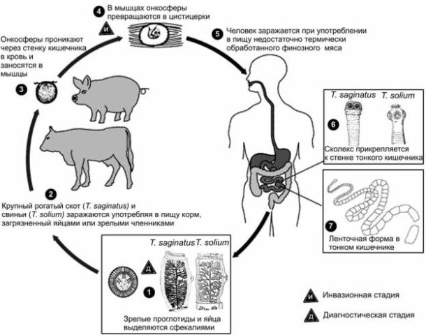
Life cycle of the bovine tapeworm, including bovine tapeworm disease and human teniarinosis
Types of fynosis in cattle
Strictly speaking, there is only one type of bovine fynosis: the one caused by Cysticercus bovis, the larva of Taeniarhynchus saginatus/ Taenia saginata (in this case, the Latin names are synonymous). To put it simply: fynosis in cattle is caused by the larva of the bovine tapeworm. Although, given the ultimate host of this parasite, it would be more correct to call the worm “human.”
But cysticercosis, which can affect cattle, is not limited to finnosis. Somewhat less frequently, cattle can become infected with other tapeworms. The final hosts of the tapeworm species Taenia hydatigena are carnivores, which today can rightfully include humans. In nature, scavengers become infected by eating the carcass of a dead infested animal. A person can acquire a lodger if he consumes the internal organs of farm animals.
Just like the bovine tapeworm, the carnivorous tapeworm “sows” segments into the environment. Herbivores, eating food contaminated with the excrement of predators, become infected with tenuicola cysticercosis. Animals susceptible to infection with this type of cysticercosis:
- sheep;
- goats;
- pigs;
- cattle;
- other herbivores, including wild species.
Once in the intestines, oncospheres migrate with blood to the liver, penetrate the parenchyma and enter the abdominal cavity. There, after 1-2 months, oncospheres turn into invasive cysticerci.
Tenuicol cysticercosis differs from bovine fynosis in that it is distributed almost everywhere. It does not have areas of maximum distribution, like Finnose. The only thing that helps is that cattle are infected with tenuicol cysticercosis less often than with finnosis.
Another type of cysticercosis is “cellulose”, also called finnosis. But Taeniasolium larvae do not parasitize cattle. They amaze:
- cats;
- bears;
- pigs;
- dogs;
- camels;
- rabbits;
- person.
Cysticercosis caused by Cysticercus cellulosae is also called porcine finnosis. For pork tapeworm, humans are both the intermediate and the final host. If we get lucky".
These diseases are simply called differently. And the intermediate hosts of other cestodes are different.
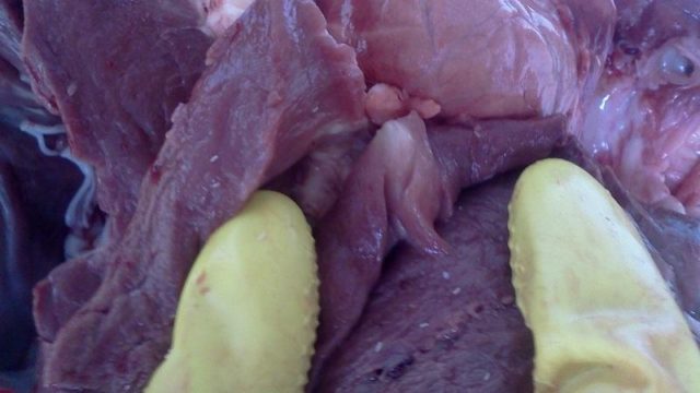
When carefully cutting up a cattle carcass infected with finnosis, you can see cysticerci. These are the white spots in the photo
Symptoms of bovine fynosis
The manifestation of clinical signs of cysticercosis depends on the degree of infection. If it is mild, the animal may not show symptoms at all. When cattle are heavily infected with cysticercosis bovis, the following is observed:
- increased body temperature;
- weakness;
- muscle tremors;
- oppression;
- lack of appetite;
- rapid breathing;
- intestinal atony;
- diarrhea;
- moans.
These signs last for the first 2 weeks, while the larvae migrate from the intestines to the muscles. Then the symptoms of Finnosis disappear and the animal “recovers.” The owner is happy that everything worked out.
Signs of infection with cysticercosis tenuicola are noticeable only during the acute course of the disease, while the larvae migrate through the liver to the site of localization:
- heat;
- refusal of food;
- frequent heartbeat and breathing;
- anxiety;
- icteric mucous membranes;
- anemia;
- diarrhea.
With severe infection with tenuicol cysticercosis, young animals may die within 1-2 weeks. The disease then enters the chronic stage and proceeds with uncharacteristic symptoms or asymptomatically.
Diagnosis of cysticercosis in cattle
A lifetime diagnosis of cysticercosis in cattle is made using immunological methods. But it is possible to determine exactly what type of cysticercosis an animal is suffering from only posthumously.
The diagnosis is usually made only after the animal is slaughtered. In bovine cysticercosis, the larvae are localized in the striated muscles. Simply put, in the same beef that comes to the table in the form of steaks, entrecote and other delicacies. True, you have to be very careless to start cooking this meat. If cattle are infected with cysticercosis, there is no need to examine the meat under a microscope: the diameter of the bubbles located between the muscle fibers is 5-9 mm.
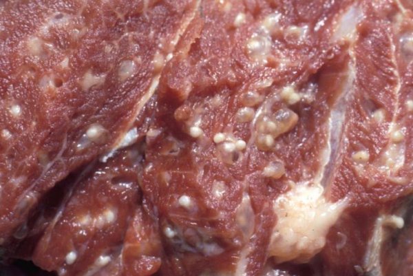
This is what the meat of an animal infected with bovine cysticercosis looks like in the photo.
They are clearly visible to the naked eye. But you can play natural scientist, take a microscope and admire the double membrane and one scolex of the cysticercus that causes bovine finnosis.
When the carnivorous worm Taenia hydatigena is infected with cysticerci, the larvae are even more difficult to miss. Cysticercus tenuicollis are localized in internal cavities and organs and are the size of a chicken egg. And if you want it, you won’t miss it.
In the acute course of cysticercosis tenuicola, changes in the internal organs are detected in dead young animals:
- the enlarged liver has a clayey color;
- on the surface of the liver there are pinpoint hemorrhages and tortuous bloody passages in the parenchyma;
- in the abdominal cavity there is a bloody fluid in which fibrin and small translucent white bubbles float.
These vesicles are the migrating cysticerci of carnivorous tapeworms. When washing the crushed liver, young larvae are also found.
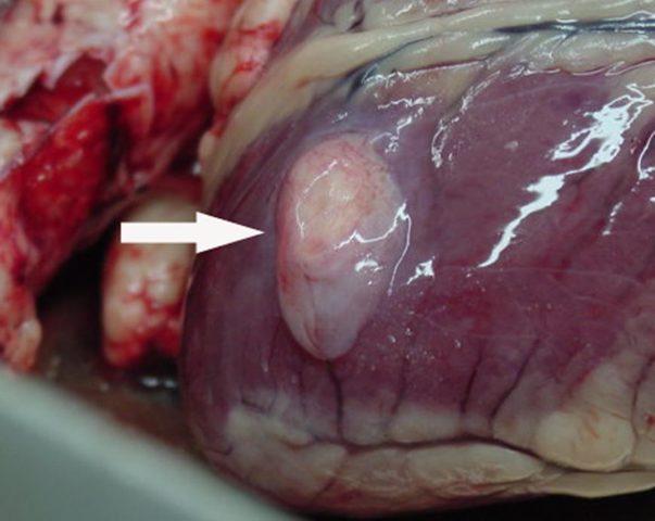
Cysticercus tenuicollis in cardiac muscle
Treatment of cysticercosis in cattle
Until recently, all reference books indicated that treatment for Finnoses has not been developed, since the larvae in cysticerci (sphere capsules) are well protected from the action of anthelmintic drugs. Sick cattle are slaughtered and the meat is sent for deep processing. I mean, they make meat and bone meal from carcasses, which is then used as fertilizer and animal feed.
Today, bovine fynosis is treated with praziquantel. The dosage is 50 mg/kg live weight. Praziquantel is administered over 2 days. The drug can be injected or added to food. The manufacturer of the drug is the German company Bayer. But we must take into account that complete confidence in curing an animal from bovine fynosis can only be obtained after slaughter and examination of the cysticerci under a microscope (live or dead).
However, for the owner of dairy cattle, only the acute stage of bovine fynosis poses a danger, when the larvae migrate into the muscles. At this time, cysticerci can also enter the milk ducts. Later, it will no longer be possible to become infected through milk.
Preventive actions
Prevention of cysticercosis in cattle has to be carried out not only on the farm where the infection was discovered, but throughout the entire region. Household slaughter of animals is prohibited. All cattle meat that comes from farms and settlements in contaminated areas is carefully monitored. Restrict the movement of stray animals. Simply put, stray dogs are shot, and their owners are required to be put on a chain.
Animals sent for slaughter are tagged to identify foci of infection with finnosis and to identify people with teniarinhoz. Carcasses infected with cysticercosis are rendered harmless following veterinary and sanitary rules.
Farm personnel are examined quarterly for infection with teniarinhoz. People found to have tapeworm are removed from serving animals.
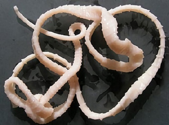
Uncooked meat from an animal with Finnosis is the source of this cute pink worm appearing in the human gastrointestinal tract
Threat to humans
A cysticercus that enters the human body along with uncooked cattle meat quickly turns into a young tapeworm. The worm grows and after 3 months begins to shed its mature segments.
It is “unprofitable” for the parasite to be quickly detected, and the most common sign of infection with teniarhynchosis is the release of these same segments. The “capsules” may appear to be separate organisms because they show some of the characteristics of small flatworms: they crawl. The patient also feels itching in the anus.
Due to the fact that the animal inside is already large, the patient may experience:
- nausea and vomiting;
- attacks of abdominal pain;
- increased appetite with weight loss;
- sometimes appetite decreases;
- weakness;
- Digestive disorders: diarrhea or constipation.
Sometimes signs of allergies are noted. Few people associate other signs with helminthic infestation:
- blood from the nose;
- dyspnea;
- heartbeat;
- noise in ears;
- flickering black dots before the eyes;
- discomfort in the heart area.
With multiple infections with bovine tapeworm, dynamic intestinal obstruction, cholecystitis, internal abscesses, and appendicitis are noted.
The discarded segments, exhibiting considerable mobility, can enter through the Eustachian tube into the middle ear or into the respiratory tract. To do this, they first need to get into the oral cavity, which they do by excreting themselves in vomit.
Pregnant women infected with bovine tapeworm may:
- spontaneous abortion;
- anemia;
- toxicosis;
- premature birth.
This “cute and very useful for weight loss” worm can start in a person:
Conclusion
Finnosis in cattle is not as dangerous to the animals themselves as to humans. It is almost impossible to remove larvae from muscle fibers. Even after the application of praziquantel and the death of the larvae, the spheres themselves will remain in the muscles.
