Content
Ungulates are phalange-walking animals. This means that the entire weight of their body falls on only a very small point of support - the terminal phalanx on the fingers. The keratinized part of the skin: nails in humans, claws in many mammals and birds, in ungulates in the process of evolution it turned into a hoof. The outer part of this organ bears at least half of the total load on the entire hoof. Because of this, hoof diseases in cattle and horses are very common. Sheep, goats and pigs also suffer from hoof diseases, but to a lesser extent because they weigh less.
Types of hoof diseases in cows
The hoof is a horny capsule that protects the tissues inside, firmly connected to the skin. The structure of a cow's hoof is similar to that of a horse. The only difference is that cows have two fingers.Because of this, a cow's hoof wall is slightly thinner than a horse's. The soft part of the sole also has a slightly different shape. But the principle is the same.
The hoof is not a monolith. It has a complex structure. The hard part of the hoof, called the hoof shoe, consists of the following layers:
- The hoof wall formed by the tubular horn. This part is “dead” along almost the entire height of the hoof and performs a protective function.
- Leaf horn located under the tubular layer. This layer, closer to the plantar part, also dies off and forms a “white line”: a relatively soft substance resembling rubber. The leaf layer is “alive” along almost the entire height of the hoof, except for the plantar part.
- The sole protects the hoof underneath.
The dead and hard layers of the hoof are separated by the living layers of skin that surround the coffin bone on the sides and bottom.
Inside the hoof shoe are the bones of the two phalanges of the toe. Cows walk on the terminal phalanx, which is called the hoof bone. The hoof shoe follows the shape of this bone.
The hoof shoe is connected to the skin of the limb through a special layer: the skin of the corolla. The width of the corolla is only about 1 cm. But this area plays an important role in the formation of the hoof. Damage or disease to the corolla also affects the hooves of cattle.
The most common fungal diseases in cows are:
- Mortellaro disease;
- pododermatitis;
- footrot.
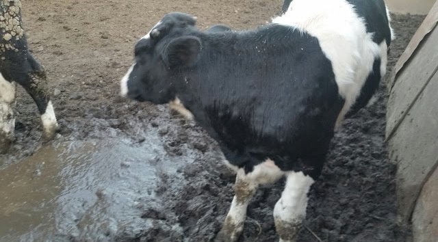
Favorable conditions for the development of various types of fungi are created by dirty litter and insufficient exercise.
This “injustice” is explained by the fact that it is often more profitable to sell a cow for meat than to spend money on treating the disease. The same techniques are used for especially valuable breeding cows as for horses.
Strawberry disease
The popular name for digital dermatitis. This disease has synonyms associated with the author of the discovery and the place of first discovery:
- hairy heel warts;
- strawberry foot rot;
- Mortellaro disease;
- Italian rot;
- papillomatous digital dermatitis.
All disease names reflect either the history of discovery or the appearance that the skin lesion takes.
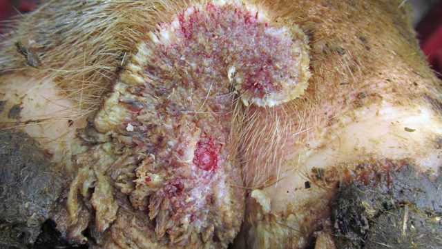
Digital dermatitis was first discovered in Italy (Italian rot) in 1974. The disease is caused by mixed types of bacteria, instead of one specific pathogen. Externally, the affected area looks like a pink tumor with tubercles. A hair sticks out from each tubercle. Hence the main popular names for dermatitis: strawberry and hair dermatitis.
The real heel, similar to humans, in animals is located next to the hock joint and is called the calcaneal tubercle.
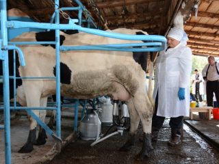
Digital dermatitis is different from foot rot, although both diseases can occur at the same time. The development of Mortellaro disease begins with damage to the heel of the hoof. The disease affects dairy cattle. Due to pain and discomfort, the cow reduces milk yield, but the quality of the milk does not suffer.
Causes and symptoms
This type of disease does not have a pronounced seasonality, since the bacteria multiply in the dirty bedding of the barn. The causes of Mortellaro disease are non-compliance with the rules of caring for cows:
- dirty wet litter;
- lack of hoof care;
- unbalanced diet, which reduces immunity;
- soft hooves;
- introduction of sick animals into the herd.
This type of dermatitis is caused by anaerobic bacteria, for which dirt in the litter is an ideal breeding ground. The basis of the “set” of bacteria are spirochetes of the genus Treponema.
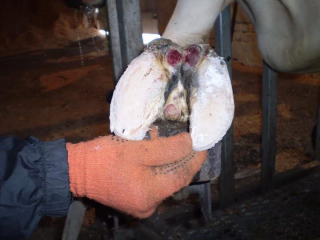
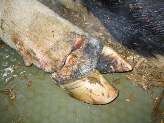
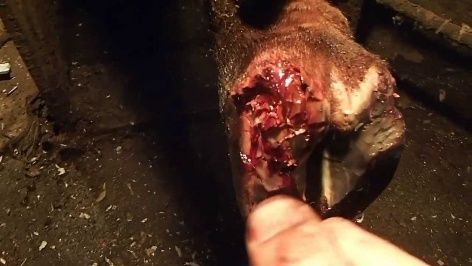
At the initial stage of the disease, the formation looks like an oval, red, raw ulcer on the heel. Then the ulcer develops into a convex lump, the surface of which resembles not the well-known strawberry, but a lychee with hairs protruding from the tubercles. But few people have seen lychees.
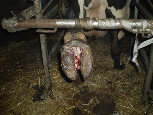
Without treatment, dermatitis grows and invades nearby areas. The formation can pass into the gap between the hooves and further upward. With advanced dermatitis, lameness is observed in the cow.
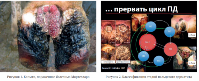
Attempts to identify the existing set of bacteria are made very rarely, and diagnosis is made based on history and clinical signs. A classification of the stages of digital dermatitis has been developed. The letter "M" in the stage designation stands for "Mortellaro":
- M0 – healthy skin;
- M1 – early stage, lesion diameter <2 cm;
- M2 – active acute ulcer;
- M3 – healing, the affected area is covered with a scab;
- M4 – chronic stage, most often expressed as thickened epithelium.
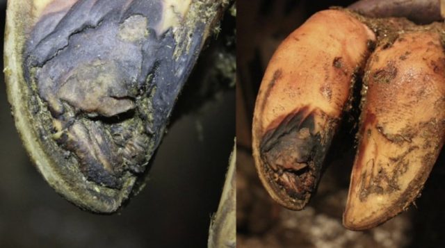
For digital dermatitis, comprehensive treatment is carried out, aimed at maximizing the destruction of all possible types of pathogenic bacteria.
Photo of a cow's hoof with Mortellaro disease and its development cycles.
Treatment methods
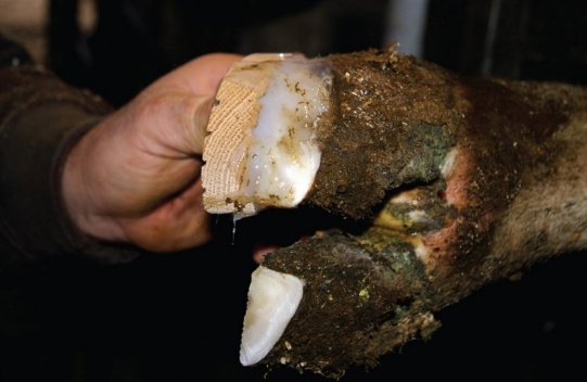
Treatment of the disease is carried out with the help of antibiotics, which are applied to the affected areas. The skin must first be cleaned and dried.The best treatment for Mortellaro disease is Oxytetracycline, which is applied to the ulcer. Dressings do not affect the course of treatment, but protect the wound from contamination. This procedure is done at will.
If many animals in the herd are sick, take baths with a disinfectant solution. The solution contains formaldehyde and copper sulfate. The second option is thymol solution.
The length of the bath is at least 1.8 m, and the depth is at least 15 cm. It is made in such a way that each leg of the cow is dipped twice in the solution to the level of the fetlock joint. The barn avoids the formation of slurry, which promotes the development of pathogenic bacteria.
Footrot
Also a multibacterial hoof disease, but the predominant microorganisms causing the rot are Fusobacterium necrophorum and Bacteroides melaninogenicus. Foot rot affects cattle of any age, but is most common in adult cows.
The disease does not have a pronounced seasonality, but in rainy summer and autumn, cases of the disease become more frequent.
Causes and symptoms
If the skin is healthy, bacteria cannot cause disease. To penetrate the body, pathogens need some kind of damage to the skin. Provoking factors are:
- Dirt and wet bedding soften the skin. Because of this, the epidermis is easily damaged, and infection can penetrate through the wound.
- Mud that has frozen into sharp spikes or dried to a hard state can also injure a cow's leg.
- Stones often injure the skin around the hoof.
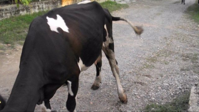
Since it is difficult to injure all 4 legs at the same time, usually the symptoms of the disease appear first on one limb.
Signs of the initial stage of the disease:
- lameness;
- wound damage on the sore leg;
- pus may be present;
- unpleasant odor;
- fever with temperature 39.5-40 °C;
- leg swelling;
- acute pain.
Typically, foot rot is a difficult to treat cattle hoof disease, and treatment can take several months. Especially under poor conditions. But there have also been cases of spontaneous recovery.
Treatment methods
In the case of foot rot, you shouldn’t hope for it to “go away on its own.” Typically, this disease is well treated with systemic antibiotics in combination with preventive measures: dry, clean bedding and long periods of walking on pasture.
Among the antibiotics used to treat the disease are:
- tetracyclines;
- penicillin;
- sodium sulfadimidine;
- sulfabrommetazine;
- other antibacterial agents.
After treatment with medications, cows are kept on a clean, dry floor until signs of rot disappear.
Recent studies abroad have shown the high effectiveness of zinc supplements in disease prevention. Also, as a preventive measure, chlortetracycline is added to livestock feed at the rate of 2 mg per 1 kg of live weight.
Pododermatitis
Pododermatitis is a group of diseases:
- aseptic (non-purulent or non-infectious);
- infectious (purulent);
- chronic verrucous.
The causes and symptoms of these cow hoof diseases, as well as their treatment, differ from each other.
Aseptic pododermatitis
This is a non-purulent inflammation of the underlying skin of the hoof.The disease has 2 types of progression: acute and chronic. Pododermatitis can be localized to a limited area or involve a significant portion of the hoof. The most common site of occurrence of the disease is the area of the heel corners.
Causes and symptoms
There are quite a few reasons for the occurrence of non-purulent pododermatitis, but usually they are all associated with excess pressure on the sole:
- bruises (in simple terms they are often called bruises);
- improper hoof trimming, due to which the cow begins to rely not on the hoof wall, but only on the sole;
- thinning of the sole due to improper trimming;
- maintenance and movement on a solid surface.
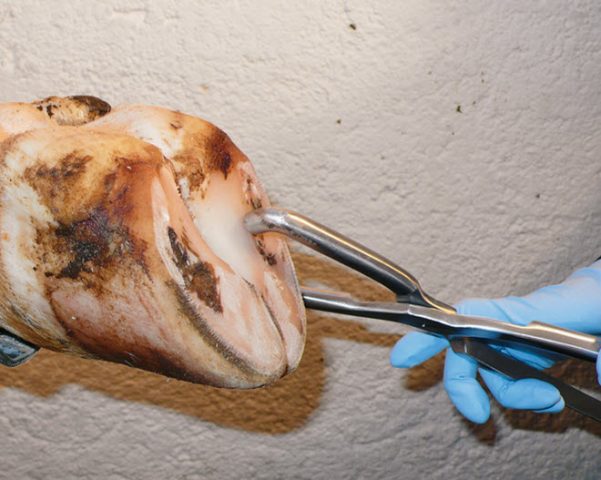
The symptom of this type of disease is lameness, the degree of which depends on the severity of the damage to the hoof. In acute aseptic pododermatitis, lameness intensifies when moving on hard ground. The temperature of the hoof shoe is higher than on a healthy limb. This difference is determined by simply feeling with your hand. Increased pulsation of the digital arteries. The localization of inflammation is determined using test forceps.
The chronic form of the disease is determined by the appearance of the hoof.
Treatment methods
The cow is transferred to soft bedding. On the first day, cold compresses are applied to the hoof. From the 2nd day until the end of the inflammation process, thermal procedures are used: hot baths or mud, UHF.
Injection of corticosteroids into the digital arteries is also recommended. But this procedure must be carried out by a specialist.
If the inflammation does not go away or the symptoms worsen, the abscess is opened. The open cavity is protected with a sterile dressing until scarring occurs.
Chronic aseptic pododermatitis in cows is not treated, since it is not economically profitable.
Infectious pododermatitis
The disease occurs in all species of ungulates. The flow can be superficial or deep; diffuse or focal.
Causes and symptoms
The cause of the disease is usually infection of wounds, deep cracks and swellings. In cows, infectious pododermatitis often occurs as a result of prolonged confinement on hard cement floors. In this case, the occurrence of the disease contributes to the abrasion and softening of the sole of the hoof.
The main sign of purulent pododermatitis in a cow is protecting the leg. The cow at rest rests only on the toe of the affected leg. Lameness is clearly visible when moving. The general temperature of cows increases slightly, but the hoof is hot to the touch. When examined with test forceps, the cow pulls out her leg and does not want to stand still.
With deep purulent pododermatitis, the signs of the disease are the same as with superficial ones, but more pronounced. If the lesion has not yet been opened, the general depressed state of the cow is also observed.
Treatment methods
When treating a disease, the abscess is first opened, since it is necessary to ensure free passage of pus. The source of inflammation is detected using test forceps and then the sole is cut out until the abscess is opened.
After the operation, the wound is washed with an antiseptic from a syringe, dried with cotton swabs and then treated with antibacterial powder preparations. A sterile bandage is applied over it. If the lesion was opened from the plantar side, the bandage is soaked in tar and a canvas stocking is put on.
Chronic verrucous pododermatitis
The old name of the disease is “arrow cancer.”Previously it was believed that this hoof disease was unique to horses. Later, verrucous pododermatitis was discovered in cows, sheep and pigs. The disease usually affects 1-2 fingers, rarely when all the hooves on a limb are damaged.
Cancer of the frog begins from the crumb, less often from the sole of the hoof. This type of dermatitis received the name “arrow cancer” because the tissues damaged by the disease look like neoplasms.
Causes and symptoms
The causative agent of the disease has not been identified. Provoking factors include:
- keeping in dirt;
- prolonged softening of the hoof horn due to damp soil;
- excessive cutting of the finger crumb.
In the benign form of the disease, hyperplasia of the papillary layer is present. In the malignant form, histology studies show carcinoma.
Hyperplasia and breakdown of the stratum corneum are detected from the moment clinical signs of the disease appear. The papillae of the base of the stratum corneum, increasing, take on a flask-shaped shape.
In the affected area, the stratum corneum becomes soft, begins to separate easily and turns into a liquid brown mass with an unpleasant odor. Gradually the process spreads to the entire pulp and sole of the hoof. The process does not affect the stratum corneum of the hoof shoe, but secondary purulent abscesses occur in this area of the hoof, as well as in the area of the corolla and spinal cartilages.
Lameness is most often absent and only appears when walking on soft ground or when the hoof is severely damaged.
Treatment methods
No effective treatments have been found for this disease. The affected areas are cut out and then cauterized with antiseptic agents. A positive result is obtained if the disease was in the initial stage. In severe cases, it is more profitable to sell the cow for meat.
Laminitis
This disease also belongs to the group of pododermatitis. Since the mechanism of occurrence and course of the disease differs from other types of diseases in this group, laminitis is usually not perceived as pododermatitis. The common name for this disease is “opoy”. But modern research has proven that water is not a factor causing this disease. Moreover, the name “opoy” comes from the fact that the disease allegedly occurred due to a hot horse drinking a large amount of water. But cows, sheep and goats also suffer from laminitis. But no one chases these animals until they are exhausted.
Laminitis has other names:
- rheumatic inflammation of the hooves;
- acute diffuse aseptic pododermatitis.
Horses are indeed the most susceptible to the disease. In all species of ungulates, the disease most often affects the forelimbs due to the fact that the main weight of the animal falls on the shoulder girdle. Less commonly, all four legs may be affected.
Causes and symptoms
Unlike other pododermatitis, rheumatic inflammation of the hooves is toxic and chemical in nature. The causes of the disease are:
- protein-rich food for lack of exercise;
- low-quality moldy feed contaminated with fungal toxins;
- excess weight;
- keeping on a hard floor;
- tympany;
- infectious diseases;
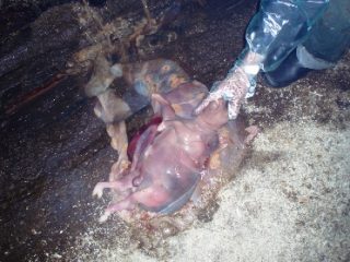
- postpartum complications;
- abortions;
- a dead fetus decomposing in the uterus;
- allergies to medications.
The first signs of the disease are easy to miss, since only in the first hours are rapid breathing, an increase in general body temperature, and cardiac disturbances observed. At the same time, muscle tremors and hyperemia of the mucous membranes appear. These signs can be confused with many other diseases.
Afterwards, the body temperature returns to normal, breathing and heart function are restored. Externally. Since the cow has an unnatural stance with the hooves resting on the heel. When listening, you will notice a rapid heartbeat: a sign of pain.
Rheumatic inflammation of the hooves can occur in two forms: acute and chronic. In acute inflammation, hoof pain increases during the first 2 days. Later, the pain subsides, and after a week, complete recovery may occur. But in fact, if left untreated, acute hoof inflammation often becomes chronic.
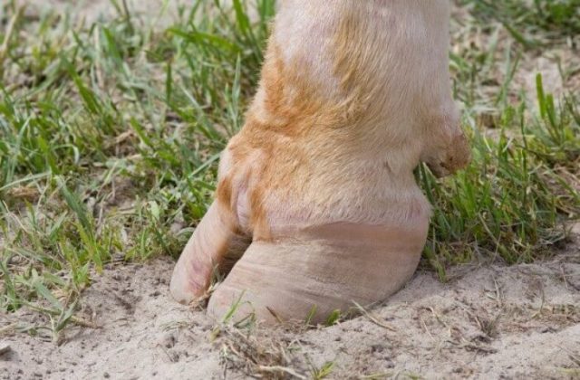
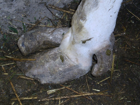
In the chronic form of the disease, the coffin bone becomes displaced and, in severe cases, protrudes through the sole (sole perforation). The hoof becomes “hedgehog-like”. Well-defined “waves” of the hoof horn appear on the front of the hoof. This is due to the fact that the toe part of the hoof during rheumatic inflammation grows much faster than the heel part.
If the disease is particularly severe, the hoof shoe may come off the limb. For any ungulate animal, this is a death sentence. If they try to treat horses as pets, then saving a cow makes no sense. It's cheaper to buy a new one. Most often, the shoe comes off from only one hoof. Since a cow is a cloven-hoofed animal, it has a chance to survive if the shoe comes off only one hoof on its leg. But, in essence, the cow will remain mutilated.
The horse was even saved, spending a lot of time and money. But he was no longer fit for work.
Treatment methods
If the hoof is deformed, treatment is no longer possible.A favorable prognosis for the outcome of the disease is only possible if measures are taken within the first 12-36 hours.
First of all, the cause of the disease is removed. The cow is transferred to a box with soft bedding. Cooling wet compresses are applied to the hooves. A good option is to place the cow in a stream so that the hooves can be cooled by the running water. Analgesics are used to relieve pain. Emergency weight loss in a cow, although not very significant, can be achieved by giving diuretics. Losing weight is necessary to reduce pressure on the hooves. Once signs of acute inflammation have resolved, the cow is moved to improve blood circulation in the hooves.
Cellulitis of the corolla
Purulent inflammation of the fiber located under the base of the skin of the corolla and hoof border. There are two types of phlegmon: traumatic and infectious. The first occurs when the skin of the corolla is injured or severely softened. The second is a complication of other hoof diseases.
Causes and symptoms
The cause of the disease is most often repeated bruises and wounds of the corolla. When kept on dirty bedding for a long time, the skin of the corolla softens, and microorganisms that cause disease can also penetrate through it. Moments that contribute to the appearance of purulent inflammation of the hoof: low immunity in the cow due to exhaustion, overwork or another disease. Cellulitis can also be a consequence of purulent-necrotic processes in the cow’s hoof.
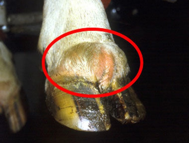
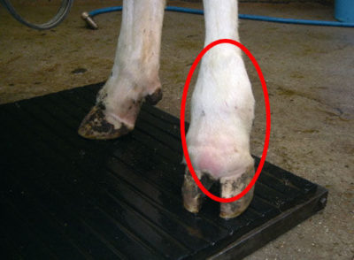
The first sign of the onset of the disease is swelling of the corolla of the hoof with an increase in local temperature. The swelling is painful and tense. A little later, other symptoms of the disease appear:
- increase in general body temperature;
- decreased appetite;
- oppression;
- decrease in milk yield;
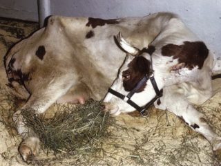
- severe lameness;
- reluctance to move, the cow prefers to lie down.
A blood test may show too many white blood cells in a cow's blood.
With further development, the tumor increases and hangs over the hoof wall. Swelling spreads to the entire finger. At the highest point of the tumor, softening appears and the skin tears, releasing the accumulated pus. After opening the abscess, the cow's general condition immediately improves.
With the second type of phlegmon (purulent-putrefactive), first a whitish stripe appears on the lower edge of the swelling. On the 3-4th day, brownish drops of exudate appear on the surface of the swelling. On the 4-5th day, the skin becomes necrotic, the exudate becomes bloody, and ulcers appear in place of the torn pieces of skin.
In cows that have recovered from phlegmon, changes occur in the papillary layer of the corolla. As a result, even after recovery, visible defects remain on the horny wall of the hoof.
Treatment methods
The method of treatment is chosen depending on the degree of development of phlegmon and the complexity of the ongoing purulent-necrotic processes. At the initial stage of the disease, they try to stop the development of an abscess in the hoof. For this purpose, alcohol-ichthyol dressings are used. Also, antibiotics with novocaine are injected into the arteries of the cow's finger.
If the development of phlegmon has not stopped, the abscess is opened. The opening of the abscess and further treatment of the wound should be carried out by a specialist, since inflammation can already spread to neighboring tissues. The wound in the hoof is washed with hydrogen peroxide, dried and generously sprinkled with tricillin or oxytetracycline powder mixed with sulfadimezine. A sterile bandage is applied on top, which is changed every 3-6 days. In parallel with the treatment of the wound, the cow is given general tonics.
Sole ulcer
Cows do not have such a disease as hoof erosion, but a specific sole ulcer most closely matches this name. It is observed in cows in large industrial complexes. Large cows of high-milk breeds usually get sick when they are kept in stalls for a long time and are fed heavily. The disease almost never occurs in bulls. Young cattle are also less susceptible to this disease.
Causes and symptoms
Most often the disease begins in the rear hooves of the cow. Provoking factors are:
- slatted floors;
- short, cramped stalls;
- untimely hoof trimming.
When trimmed infrequently, the cow's hooves take on an elongated shape. Because of this, the balance of the cow's body is shifted, and the coffin bone takes an unnatural position.
Symptoms may vary depending on the severity of the disease:
- careful movements;
- lameness when supporting the leg, especially pronounced when moving on an uneven surface;
- the cow prefers to lie down;
- appetite decreases;
- observe gradual exhaustion;
- Milk yield decreases.
In the initial stage of the disease, gray-yellow, red-yellow or dark red spots form on the sole of the hoof. At this point the horn loses its elasticity and strength. As a result of gradual chipping of the sole, a purulent-necrotic ulcer is formed at the site of the outbreak.
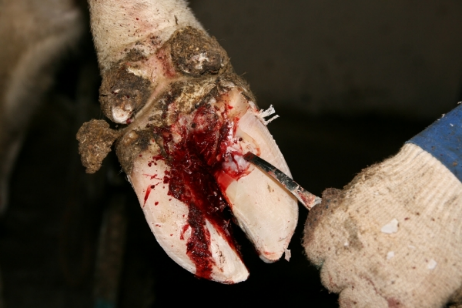
In the middle of the ulcer there is dead tissue, along the edges there are granulation growths. In case of necrosis and rupture of the deep digital flexor muscle, a fistula with a depth of more than 1 cm is formed in the ulcer. The cow lifts her leg onto the toe at the moment of support on the floor.Damage to the shuttle mucous bursa or claw joint is indicated by the flow of viscous fluid from the fistula.
Treatment methods
Hoof treatment is carried out surgically. The prognosis is favorable only at the initial stage of the disease. During the operation, all changed hoof horn and dead tissue are removed. Sometimes amputation of the affected finger may be necessary.
Tiloma
Another name is “limax”. Skin formation. This is a dense cushion in the area of the arch of the interhoof fissure.
Causes and symptoms
The reasons for its origin are unknown. Presumably, not only external factors, but also heredity play a role in the appearance of tiloma. This theory is supported by the fact that tiloma most often occurs in cows under 6 years of age. In cows older than this age, the disease is observed less frequently, and after 9 years it does not occur at all.
Signs of tiloma:
- the appearance of a dense, painless, sclerotized ridge of skin;
- the formation extends from the anterior to the posterior end of the interhoof gap;
- increase in the roller.
At the moment of support on the ground, the hooves move apart and the roller is injured. Exudate accumulates between the tyloma and the skin, irritating the skin. With repeated injuries, an infection enters the wound, leading to purulent diseases of the hoof. Sometimes the roller may become keratinized. In a cow with tiloma, first observe carefully resting the affected leg on the floor. Later lameness develops.
Treatment methods
Tyloma is usually removed surgically by cutting out the formation. Cauterizing the roller with antiseptic drugs very rarely leads to a positive result.
Lameness
Lameness is not a disease, but a symptom of emerging problems. There can be a lot of reasons for it. And often lameness is not caused by a disease of the hoof, but by a problem in the joints above.Lameness can also be caused by improper hoof development:
- thin sole;
- hoof compressed under the crown;
- crooked hoof;
- fragile and brittle horn;
- soft horn;
- cracks;
- horn column.
Some of these causes of lameness may be congenital, but they are often caused by improper and untimely hoof trimming.
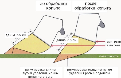
Trimming is carried out every 4 months, trying to maintain hoof balance. Trimming often takes place with adventures, since cows are usually not trained to give their feet and stand quietly during the procedure. Most often, a cow’s hoof is not paid attention to at all until the animal becomes lame. As a result, it is necessary to treat hoof diseases in cows by felling.
Prevention measures
Prevention measures for hoof diseases are simple:
- regular hoof trimming;
- keeping cows on clean bedding;
- quality walking;
- non-poisonous food;
- a lot of movement.
Prevention will not work if the disease is hereditary. But such cows are culled from the herd and are not allowed to be bred.
Conclusion
Cattle hoof diseases affect not only the movement of cows, but also their productivity. At the same time, hoof treatment is a long process and not always successful. The easiest way is to prevent illness than to correct the mistake later.