Content
Of all the invasive diseases, dictyocaulosis in cattle is the most common. Young calves are especially susceptible to infection in the autumn. If timely measures are taken, mortality in a cattle herd can be avoided, but dictyocaulosis is more difficult to cure than other invasive diseases.
What is dictyocaulosis
Parasitic worms, which are collectively called “worms,” are found not only in the gastrointestinal tract. Often a cough due to a cold is caused by a completely different reason. It is very difficult to really catch a cold. To do this you need to get very cold. But even in this case, the development of pneumonia is more likely than a “cold”.
Due to the season of infection, dictyocaulosis is often mistaken for a cold and the symptoms, rather than the cause, are treated. As a result, the disease develops and leads to the death of cattle, especially calves of the current year of birth.
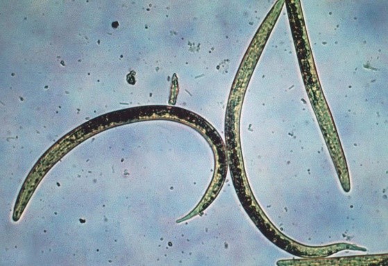
The real cause of cough in cattle is worms living in the lungs. These are nematodes: thread-like roundworms 3-15 cm long. They belong to the genus Dictyocaulus. There are several types of Dictyocaulus. Although scientists have not yet agreed on the classification of these nematodes.In cattle, the most common infection is Dictyocaulus viviparus or bovine lungworm. The same species infects wild deer and elk with dictyocaulosis. Although this is where the discrepancy lies: some scientists consider the nematode that infects wild artiodactyls to be a different species. But it has been established that in any case these parasites can cross-infect cattle and deer.
Infection of cattle with pulmonary filamentous worms is called dictyocaulosis.
Animals are generally well adapted to life in the open air. You won't get them with autumn rain.
Routes of infection with dictyocaulosis
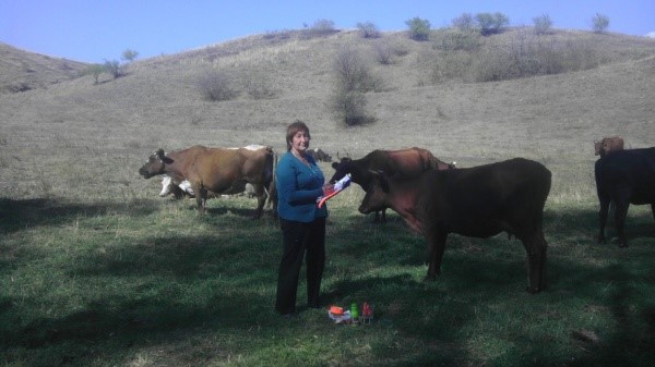
Young cattle of the first and second years of life are most susceptible to nematodes. Animals become infected with dictyocaulosis on pasture when sharing grazing with already sick individuals. Infection occurs when nematode larvae are ingested along with water or grass. The spread of dictyocaulosis in cattle is facilitated by crowded keeping of animals of different ages on pasture.
The spread of bovine dictyocaulosis on pastures is facilitated by:
- floods;
- rains;
- fungus from the genus Pilobolus.
In the southern regions, where drought is common in summer, cases of infection with bovine dictyocaulosis do not occur between July and August. In central Russia, the “disease season” lasts from spring to autumn.
Life cycle of Dictyocaulos
Parasites have a simple but very interesting life cycle, as they spread using mold. Adult nematodes live in the branched passages of the bronchi. This is where they lay their eggs.Since the worms irritate the bronchi as they move, cattle cough reflexively. The laid eggs are “coughed up” into the oral cavity, and the animal swallows them.
The first stage larva (L1) emerges from the eggs into the gastrointestinal tract. Next, the larvae, along with the host's manure, enter the environment and develop in feces during the next two stages.
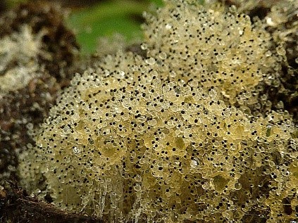
A mold fungus of the genus Pilobolus grows on manure. In the L3 stage, the larvae penetrate the fungi and remain there, in the sporangia (organs in which spores are formed), until the fungi mature. When a mature fungus releases spores, the larvae fly away along with them. The radius of dispersal of larvae is 1.5 m.
Pilobolus spores pass through the intestines of cattle and in this way can spread over considerable distances.
In the wild, animals don't eat grass near their own species' feces, but in grasslands they have no choice. Therefore, along with grass, cattle also swallow L3 stage larvae.
Parasites enter the gastrointestinal tract of cattle and pass through the intestinal wall, entering the lymphatic system of cattle and through it they reach the mesenteric lymph nodes. At the nodes, the larvae develop to the L4 stage. Using the bloodstream and lymphatic system, L4 enter the animal's lungs, where they complete development, becoming adult nematodes.
Symptoms of dictyocaulosis in cattle
Signs of bovine dictyocaulosis are often confused with a cold or bronchitis. As a result, dictyocaulosis in cattle enters a severe stage and leads to death. Calves are especially affected by dictyocaulosis. The picture of the disease is not always clear, as it largely depends on the general condition of the animal. But usually there are:
- oppression;
- cough;
- elevated temperature;
- shortness of breath when inhaling;
- rapid breathing;
- rapid pulse;
- serous discharge from the nostrils;
- exhaustion;
- diarrhea;
- tactile fritmit.
The latter means that the vibration of the lungs during breathing in cattle can be “felt” through the ribs.
In advanced cases, dictyocaulosis is complicated by pneumonia, drags on for a long time and ultimately leads to the death of cattle. When dictyocaulosis passes into the terminal stage, the animal will not live long:
- attacks of severe painful cough;
- constantly open mouth;
- a large amount of foam from the mouth;
- heavy breathing, wheezing.
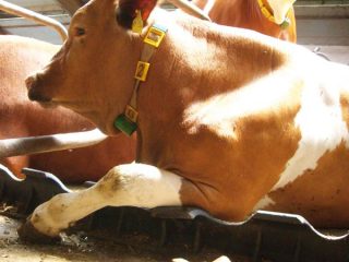
Due to the lack of air in the lungs clogged with worms, the cow suffocates: she falls on her side and lies motionless, not reacting to external stimuli. This stage of dictyocaulosis quickly ends with the death of the animal.
Diagnosis of dictyocaulosis in cattle
A lifetime diagnosis of “dictyocaulosis” is established taking into account epizootological data, the general clinical picture and the results of analyzes of cattle feces and sputum coughed up by animals. If nematode larvae are found in manure and pulmonary secretions, there is no doubt that the cough is caused by dictyocaulosis pathogens.
Nematodes are different. Many of them live freely in the soil and feed on decaying organic matter. Such worms can also crawl to manure lying on the ground. But the presence of L1 stage larvae in rectal manure is a sure sign of dictyocaulosis in cattle.
Pathological changes in dictyocaulosis in cattle
In a dead animal, a pathological examination reveals catarrhal or purulent-catarrhal pneumonia and a foamy mass in the bronchi. The latter is precisely the habitat of adult parasites.
The walls of the blood vessels in the lungs are hyperemic.The affected lobes are dense, enlarged, dark red. Mucous membranes are swollen. Areas of atelectasis are noticeable, that is, “collapse” of the alveoli, when the walls stick together.
The heart is enlarged. The wall of the heart muscle is thickened. But the option of dilatation is also possible, that is, enlargement of the heart chamber without thickening the wall. Changes in the heart muscle are due to the fact that when the lungs were clogged with worms, the animal did not receive enough oxygen. To compensate for the lack of air, the heart was forced to pump large volumes of blood.
Since the larvae from the gastrointestinal tract and mesentery “made their way” into the lungs, they also damaged the intestinal walls. Because of this, you can also notice pinpoint hemorrhages there: the places where the larvae emerge at the time of their “journey” to their permanent place of residence.
Treatment of dictyocaulosis in cattle
The main treatment for dictyocaulosis is timely deworming of cattle with special drugs that act on nematodes. But there are a lot of medicines for dictyocaulosis. There are those that have been used for more than 20 years. There are also more modern ones.
Worms are not so complex that they keep their DNA unchanged despite exposure to different substances. Therefore, like insects, they mutate and adapt to different drugs.
Old drugs:
- Nilverm (tetramizole). For cattle 10 mg/kg with feed or as a 1% aqueous solution. Set twice with an interval of 24 hours.
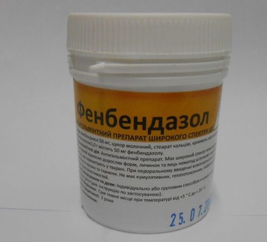
- Fenbendazole (Panakur, Sibkur, Fenkur). Dose for cattle 10 mg/kg with feed. One time.
- Febantel (rintal). For cattle 7.5 mg/kg once orally.
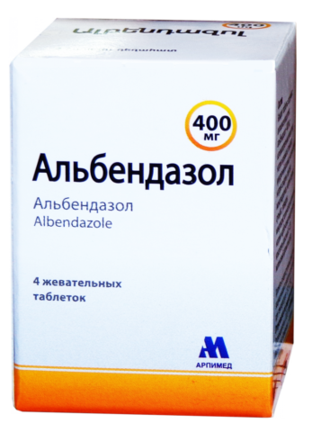
- Albendazole. 3.8 mg/kg orally.
- Mebendazole. 15 mg/kg with food.
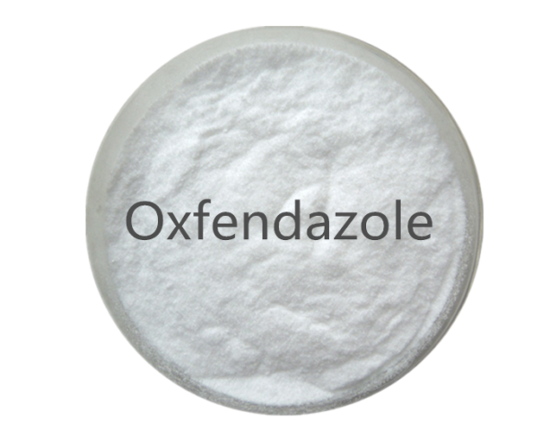
- Oxfendazole (Sistamex). 4.5 mg/kg orally.
All dosages are indicated for the active substance.
Over time, newer drugs for dictyocaulosis have appeared, which have already become familiar. Some of them are complex, that is, they contain more than one active ingredient:
- Levamectin: ivermectin and levamisole. 0.4-0.6 ml/10 kg. Used for dictyocaulosis of heifers;
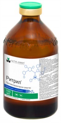
- Ritril. Used for the treatment of young cattle. Dose 0.8 ml/10 kg, intramuscular.
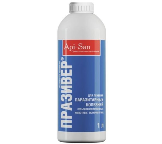
- Praziver, active ingredient ivermectin. 0.2 mg/kg.
- Monesin. For adult cattle 0.7 ml/10 kg orally, once.
- Ivomek. For young cattle 0.2 mg/kg.
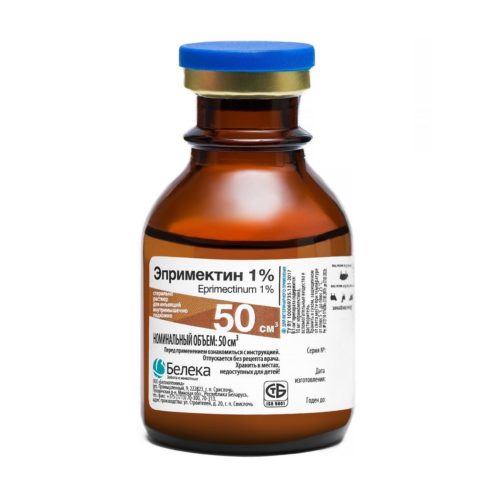
- Eprimectin 1%.
The latter drug has not yet been licensed, but recovery of cattle from dictyocaulosis after its use was 100%. The drug is produced in Belarus. Complete liberation of cattle from nematodes occurs already on the fifth day after using the new generation of drugs. Today, in the treatment of dictyocaulosis, aversectin anthelmintics are already recommended.
Treating calves the old fashioned way
Nematodes are driven out of the lungs of cattle using the “miraculous” iodine. This method is used in relation to calves, which are easier to kill than an adult.
Preparation of the solution:
- crystalline iodine 1 g;
- potassium iodide 1.5 g;
- distilled water 1 l.
Iodine and potassium are diluted in water in a glass container. The calf is rolled over and placed in a dorsolateral position at an angle of 25-30°. Dose per lung 0.6 ml/kg. For therapeutic purposes, the solution is injected into the trachea using a syringe, first into one lung, and a day later into the other. For preventive purposes - into both lungs at the same time.
Preventive actions
Considering that it is very difficult to remove nematodes from the lungs, and that dead worms begin to decompose there, prevention is more cost-effective.To prevent infection with dictyocaulosis, isolated housing of calves is practiced:
- stall;
- stall-camp;
- stall-walking;
- grazing in areas free from grazing since last fall.
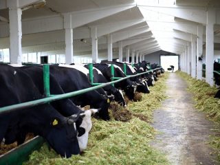
Calves are separated by age groups so that older and possibly infected individuals do not transmit nematodes to the young.
On pastures, young cattle are regularly examined for dictyocaulosis (manure analysis). Surveys begin one and a half months after the start of grazing and are repeated every 2 weeks until the end of the grazing season.
If infested individuals are found, the entire herd is dewormed and transferred to stall housing. Calves of the second year of life undergo preventive deworming in March-April. Cubs born this year are wormed in June-July. If necessary, that is, if dictyocaulus were found on the pasture, additional deworming is carried out in November before placing it in stalls.
Also, back in Soviet times, phenothiazine was fed to cattle on pasture in fractional shares, along with feed additives: salt and minerals. In areas unfavorable for dictyocaulosis, cattle are dewormed monthly as a preventive measure. But this practice is undesirable, since all anthelmintics are poisons and in large quantities poison the animal being treated.
There is one more measure that has not been adopted in Russia, but helps reduce the number of worms on pastures: regular manure removal. Since the larvae are spread by fungal spores growing on cow feces, timely removal will reduce their numbers. And along with the mold, the number of scattered larvae will also decrease.
In other words, in the West, manure is removed from pastures not because “there is nothing else to do,” but because of harsh economic considerations. Removing manure is cheaper, faster and easier than treating cattle for dictyocaulosis.
Conclusion
Dictyocaulosis in cattle can cause a lot of trouble for owners if they attribute cough and mucus from the nose to a cold. When a cow suddenly shows such signs, you first need to remember how long ago the animal received anthelmintic. And follow an important rule: when changing the housing regime, always deworm your livestock.
