Content
Dairy cattle are kept for milk. A barren cow is kept for at most 2 years: the first time barrenness could be an accident, but the animal, which has become barren, is sold for meat in the second year. But even with annual calvings, udder diseases in cows can nullify all efforts to obtain milk. Unnoticed inflammation of the udder reduces milk yield by more than half.
Types of cattle udder diseases
The types of udder diseases and their treatment in cows are not very diverse. In fact, there are only 2 diseases: edema and mastitis. But mastitis has at least 9 forms with 3 types of disease.Since one form of mastitis can turn into another if left untreated, they are not classified as separate diseases. Although some forms require specific treatment. But although the name of the udder disease is the same, in the photo the forms of mastitis look completely different.
Edema
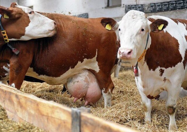
From the point of view of “classical” diseases, edema cannot even be called a disease of the udder in cows. It occurs 1.5-2 weeks before calving and is a sign of toxicosis, from which cows also suffer. That is, this is a kind of physiological reaction of the cow’s body to pregnancy. Swelling disappears 1-1.5 weeks after calving.
Causes and symptoms
Movement during pregnancy is not only for people. The main reason for udder swelling in a cow is the lack of proper exercise.
The udder is enlarged due to swelling. Smooth, while a normal udder has skin folds. When pressed, a slowly disappearing depression remains on the skin.
Treatment methods
Treatment of edema is symptomatic: massage along the lymphatic vessels from the bottom up and a laxative inside. But the easiest way is to give the cow the opportunity to move.
To prevent the disease, shortly before calving, reduce the percentage of succulent feed and increase the amount of dry feed. They make the cows walk a lot. Drink from a bucket to control the volume of water consumed.
Mastitis
Mastitis is inflammation of the udder. The forms of this udder disease in cows and their symptoms differ, depending on the cause of occurrence and the speed of decision-making on treatment. The disease can occur at any time during lactation. Sometimes a cow gets mastitis during the dry period.There are many varieties of this disease. Classification is carried out according to the nature of the inflammatory process:
- subclinical;
- serous;
- catarrhal;
- purulent;
- abscess;
- phlegmonous;
- fibrinous;
- hemorrhagic;
- gangrenous;
- specific mastitis and complications after them.
The etiology of mastitis depends on the microflora that acts as the causative agent of the disease. Bacteria can also be a factor causing complications.
Causes and symptoms
The causes of mastitis can be very diverse:
- bruises;
- wounds;
- infections;
- intoxication;
- violations of milking rules;
- poor care of the udder and milking equipment;
- rough hand milking.
Some causes of the disease overlap with each other. An uninfected wound will not cause mastitis, and it is difficult for infection to penetrate the gland if there are no cracks in the udder skin.
The causes of intoxication can also be different:
- gastrointestinal diseases;
- decomposition of the placenta retained in the uterus;
- postpartum subinvolution of the uterus;
- endometritis.
Symptoms of the disease in clinical, that is, overt, mastitis depend on the physical condition of the cow and the type of pathogen. Before treatment it is necessary to carry out a diagnosis. The main attention is paid to preventing the development of subclinical (latent) mastitis into an obvious form of the disease.
Diagnostics
Unnoticed subclinical mastitis quickly develops into an overt phase. The disease is best treated in the initial phase, before it develops into a serious problem. Diagnosis of subclinical mastitis on the farm is carried out by a veterinary specialist in the laboratory. But it is difficult for a private owner to do such research.There are 2 ways to conduct a rapid milk test for subclinical mastitis at home.
Straining
The milk is filtered through dark gauze to identify the presence of clots. If small flakes remain on the gauze after straining, mastitis is present. In the absence of illness, milk will not leave marks on the gauze.
Advocacy
10 ml of milk is poured into a test tube and kept in a regular household refrigerator for 16-18 hours. In the absence of mastitis, a layer of cream of 5 mm is formed, there is no sediment. If the cow is sick, a sediment will form at the bottom of the test tube, and the layer of cream will be thin and mixed with mucus.
Symptoms of clinical manifestations of mastitis
In addition to the types, mastitis can also have mild, moderate and severe forms. Symptoms vary depending on the form and severity of the disease. If left untreated, one type of inflammation often develops into another, more severe one.
Mild course of the disease
Subclinical, serous and catarrhal mastitis occur in a mild form. In subclinical cases there are no symptoms, but milk yield is slightly reduced.
With serous mastitis, the cow is slightly depressed and lame. Milk yield is reduced. The milk from the affected lobe is liquid with a bluish tint. Local temperatures are elevated. After milking, udder swelling does not subside. The lymph nodes on the udder are enlarged. The skin is tight and painful. In this form of the disease, the affected teats in cows are triangular in shape.
With catarrhal mastitis, the cow's condition is normal. Milk yield decreases slightly. With catarrh of the milk passages, casein clots can be seen at the beginning of milking. If catarrh has developed in the milk alveoli, clots appear at the end of milking. Local temperatures are slightly elevated. After milking, the udder “deflates.” Slight enlargement of lymph nodes.At the base of the nipple, dense cords and knots are probed. The nipple shape is oval.
Average course of the disease
Next, mastitis turns into a purulent, abscessive or phlegmonous form. It is usually difficult not to notice the disease at this stage.
With purulent mastitis, the cow is depressed and lame. There is no chewing gum. Body temperature 40 °C. There is no milk in the affected lobe. You can milk mucopurulent exudate with yellow flakes in small quantities. The lymph nodes of the udder are enlarged and painful. The skin is painful, hyperemic.
Abscess mastitis is characterized by an increase in general body temperature and refusal to feed. A reddish liquid exudate mixed with pus flows from the affected lobe. Lymph nodes are hot, painful, enlarged. Lumps or fistulas are observed on the skin.
Phlegmonous mastitis is one of the most severe forms with an “average” level of the disease. The cow is severely depressed, body temperature increased to 41 °C. There is lameness and no appetite. Secretion of the affected lobe is reduced or absent. The secretion is grayish in color, with scraps of dead tissue. With this form of the disease, the udder skin of cows is cold, doughy in consistency, and lymphatic vessels are noticeable.
Severe course of the disease
You still need to be able to survive until severe mastitis occurs. In a dairy cow, teat disease will become noticeable at the mid-stage at most. The cow will begin to kick when trying to milk her. And it is most likely that the cow will begin to shed at the beginning of the development of mastitis. Severe disease may occur in dry cows, young cows, or beef cows on large farms. Sometimes it is difficult to keep track of an individual in a large herd.Severe mastitis is expressed in fibrinous, hemorrhagic and gangrenous forms.
The fibrinous form of the disease is characterized by a depressed state of the cow, refusal to eat and lameness. The affected lobe is hot, painful, greatly enlarged, and crepitates. Discharge from the diseased nipple is straw-yellow in color with films of fibrin. With this form of the disease, the skin of the udder is thickened and hyperemic. Lymph nodes are painful, hot and enlarged.
In the hemorrhagic form of the disease, exhaustion is observed due to diarrhea. The affected part of the udder is hot, swollen and painful. There are almost no allocations. The small amount of exudate that can be milked is cloudy and watery, brown in color. Purple spots are visible on the skin of the udder. Lymph nodes are painful and enlarged.
The gangrenous form is practically untreatable. This is the final stage of mastitis development. It is characterized by sepsis, that is, “general blood poisoning” and fever. The affected lobe is cold due to cessation of blood supply. Liquid exudate with gas bubbles is released. In the gangrenous form of the disease, a smooth film forms on the skin surface of the cow's udder. Lymph nodes are very painful.
Treatment methods
Treatment of mastitis is carried out in various ways, depending on the form of the disease and the severity of its course. There are general principles for the treatment of mastitis:
- complex;
- early;
- continuous and constant;
- providing peace;
- frequent milking every 3-4 hours;
- udder massage.
To the complex treatment, which consists of increasing the cow’s immunity, specific measures are added, depending on the type of inflammation.It is necessary to start treatment as early as possible, since the inflammatory process causes the death of the alveoli that produce milk.
It is impossible to interrupt treatment until complete recovery, as the disease will return. Rest is provided to relieve tension in the mammary gland and reduce blood flow to the udder. To reduce milk production, a sick cow is transferred to dry feed and limited in water.
Udder massage is carried out according to certain patterns: for serous inflammation from bottom to top along the lymphatic channels, for catarrhal inflammation - from top to bottom from the base of the udder to the nipples.
In the first days of illness, to alleviate the condition of the cow, cold compresses are applied to the inflamed part of the udder. After 4-5 days, the inflammation enters the subacute stage, and the cold is replaced with heat. Warming compresses help resolve infiltrates. Swelling of the udder of any origin is reduced by prescribing sodium sulfate in a laxative dose once a day.
Treatment of some forms of mastitis
Mastitis accompanied by painful sensations requires specific treatment:
- serous;
- fibrinous;
- hemorrhagic;
- initial stage of abscess.
When treating these types of disease, novocaine blockades are used.
For acute mastitis with high body temperature, antibiotic therapy is used. For better effectiveness, combinations of antibiotics are used:
- penicillin + streptomycin;
- oxytetracycline + neomycin;
- ampicillin + streptomycin.
Also, when a cow’s teat is inflamed, oil-based antimicrobial drugs are injected into the milk canal.
In the final stage of treatment, mildly irritating ointments are used to absorb the remaining infiltrate.
Udder induration
This is a growth of connective tissue in the udder. Complication after mastitis or prolonged untreated swelling.
Causes and symptoms
The affected lobe is dense and does not fall off after milking. Remains large even during the dry period. Nodes may be felt in the thickness of the lobe, or it remains uniformly dense (meat udder). There is no pain.
Over time, as connective tissue grows, milk production decreases. If the process occurs in the secretory part of the mammary gland, the quality of milk deteriorates:
- gray;
- mucous;
- presence of flakes;
- unpleasant taste.
Sometimes the affected area of the udder may be smaller, then it is released with a very dense consistency.
Treatment methods
There is no treatment. Sprawl cannot be reversed.
Abscess
This is the next stage of catarrhal mastitis, which has turned into an abscess form in the absence of treatment. The photo shows the abscess stage of udder disease in a cow with an already opened abscess.
Abscess mastitis is treated.
Milk stones in the udder
A non-contagious disease that occurs due to metabolic disorders. Stones appear if phosphorus salts are deposited in the mammary gland or calcium is washed out of casein. Milk stones can also be a consequence of mastitis.
Causes and symptoms
There are only 4 reasons for the appearance of stones, but from very different areas:
- disorders in the endocrine system;
- unsanitary conditions;
- mastitis;
- incomplete milking (more often leads to mastitis than to stones).
The stones may have a clayey consistency or be hard. Their appearance is determined by palpating the nipple. It becomes hard. When palpated, compactions are detected. Stiffness also occurs.
Treatment methods
Before milking, the udder is washed with warm water and massaged from top to bottom towards the nipples. Loose stones found in the nipples can be removed using a catheter. After this, when milking, pieces of stones are removed along with the milk.
In more severe cases, all manipulations are carried out only by a veterinarian:
- surgical removal;
- destruction using ultrasound;
- course of oxytocin.
Milk is edible, but it has low fat content and high acidity. It is more suitable for making fermented milk products.
Milk incontinence
The scientific name for this phenomenon is lactorrhea. Happens quite often. But do not confuse streams of milk from an overfilled udder with lactorrhea.
Causes and symptoms
The causes of the disease may be paralysis or relaxation of the sphincter of the nipple. But problems with the sphincter also do not occur out of nowhere. The following factors can cause the cessation of the work of this muscle:
- tumor in the canal;
- mastitis;
- nipple injury;
- stressful condition.
The difference between lactorrhea and the discharge of milk from an overfilled udder is that with illness the udder may be half empty. But the milk will still drip.
Treatment is either not developed or not required. Everything will return to normal as soon as the cause that caused the sphincter to relax is eliminated.
Tightness
This is not a disease in itself, but a consequence of other problems. The most common cause of stiffness is adhesions as a result of the inflammatory process. The nipple canal narrows and stops opening.
Causes and symptoms
When the milk is tight, the milk comes out in a thin stream. The nipples harden, and when palpated, scars and adhesions may be detected. With tightness, there is a high probability that milk will remain in the udder.In this case, a vicious circle arises: mastitis-stiffness-mastitis. Sometimes the channel may close completely.
Treatment methods
At the first signs of illness, milk begins to be milked as often as possible, even if this is a painful procedure for the cow. To reduce pain, the nipples are massaged with anti-inflammatory ointment.
Bruises
A lump cannot appear on a soft udder, but a bruise can easily appear. Usually, a cow gets bruises to her udder when she is kept too crowded. When there is a conflict between cows, one may hit the other. Fresh bruises are painful and the cow may resist milking.
Treatment is limited to cold compresses for the first two days and warm compresses for the next two days. If dense areas and blood appear in the milk, you should consult a specialist. There is a very high probability that the bruise has turned into inflammation.
Cracks
Often appear during lactation due to rough milking. Through the cracks an infection enters, which leads to mastitis and furunculosis. To prevent illness, the nipples are lubricated with a moisturizing ointment. Since Soviet times, the inexpensive udder ointment “Zorka” has been popular.
Furunculosis
Bacteria penetrating through cracks in the nipples cause suppuration of the wounds, which is called furunculosis. If hygiene is not maintained, the follicles can also become inflamed.
Causes and symptoms
With the development of furunculosis, the skin of the nipples becomes rough. At the initial stage of the disease, individual foci of suppuration can be distinguished. If left untreated, the suppuration grows. The skin of the udder turns yellow-red.
Treatment methods
Treatment of the mild stage is symptomatic:
- cutting hair from the affected part of the udder;
- treating the clipped area with iodine and ichthyol ointment;
- opening mature boils and treating them with penicillin or streptocide powder, you can use an antibiotic spray.
It is advisable that the opening of boils be carried out by a specialist.
In veterinary medicine, udder diseases in cows include only edema and mastitis. The rest are either complications after mastitis, or just one of the symptoms of infectious diseases: foot and mouth disease, smallpox or nodular dermatitis. The opposite situation is also possible: mastitis is a complication of an infectious disease.
Papillomatosis
The mechanism of origin of papillomas has not been fully elucidated. They also often disappear on their own. The disease is known to be caused by a type of herpes virus. Papillomas appear when the immune system is weakened. Usually in young animals during growth.
They can also appear in an adult cow due to improper nutrition. Usually papillomas are painless, but sometimes they can cause pain. In the event that they grew near a nerve.
When milking, an external papilloma can interfere with the operation of the machine or hand. If a papilloma has grown inside the nipple, it can cause stiffness or pain.
Causes and symptoms
Very often, papillomatosis causes chronic fern poisoning, which destroys vitamin B₁. Due to vitamin deficiency, immunity is reduced, and the virus gains freedom of action.
Treatment methods
Although papillomas appear when the immune system is weakened, you cannot inject an immunostimulant at this time. Along with the body, the warts will also be “nourished”. Treatment methods are related to the prevention of the disease, since getting rid of papillomas is difficult and often impossible.
Smallpox
A viral disease contagious to mammals and birds. Characterized by fever and rash on the skin and mucous membranes.
Causes and symptoms
The virus is usually brought in from outside along with a sick cow that has not been quarantined. The incubation period of the disease is 5 days. Body temperature 41-42 °C. Skin lesions characteristic of smallpox in cows appear on the udder and teats. In bulls on the scrotum. There may also be rashes all over the body.
Cowpox is not dangerous for people, especially if they are vaccinated. Milk from a cow with smallpox is boiled or pasteurized.
Treatment methods
Only symptomatic methods are used. Pockmarks are softened with fats, and ulcers are lubricated with aseptic preparations. Antibiotics are used to prevent complications.
foot and mouth disease
A highly contagious disease that affects all mammals. It is characterized by fever and aphthae on the mucous membranes, udder skin, and in the interhoof gap.
Causes and symptoms
The causes of infection are the appearance of a sick cow in the herd or the introduction of the virus on the shoes or clothing of staff. Symptoms of foot and mouth disease are most pronounced in adult cows:
- decreased appetite;
- decrease in milk yield;
- increase in body temperature to 40-41 ° C;
- the appearance of aft.
Aphthae rupture after 12-48 hours, forming painful ulcers with ragged edges and a reddish bottom. The temperature at this point drops to normal. There is profuse drooling and lameness. After a week, the erosions heal.
With a benign course, the cow recovers after 2-3 weeks. If a complication occurs due to a secondary infection, mastitis and pododermatitis develop. With a malignant course, the cow dies after 1-2 weeks.
Treatment methods
Sick cows are transferred to a separate room and given a course of immunostimulating drugs. The mouth is washed with antiseptic drugs.The affected areas of the udder and legs are treated surgically and antibiotics, antiseptic ointments and painkillers are applied externally.
Dermatitis
There are no separate “udder dermatitis” in cows. There is an allergic reaction that can be expressed by redness and rash. It is most noticeable on the udder, as there is too little hair there. But similar signs of disease can be found throughout the cow's body.
There is a viral disease: nodular dermatitis. After the incubation period, the cow's body temperature rises. Then dense nodules appear on the skin. But also “all over the cow.” It is quite natural that these signs are most noticeable on cows with short, smooth hair or where the hair is very sparse (groin area). Lumpy dermatitis also has nothing to do with udder diseases.
Preventive actions
Almost all diseases of the udder and teats in cows come down to one type or another of mastitis. Therefore, preventive measures also refer to preventing the development of this disease. The requirements for the prevention of infectious diseases are stricter and other measures are taken in this case.
To prevent mastitis, livestock are kept in premises that meet zoohygienic requirements. The same preventive measures include providing cows with high-quality feed. If a farm practices machine milking, then all cows are selected for their suitability for this type of milking and increased resistance to udder diseases. When milking by hand, roughness is avoided: milking with a “pinch”.
One of the most important measures to prevent mastitis is the timely and correct start of cows. The launch is carried out 2 months before calving.7-10 days after launch, check the condition of the udder and the presence of fluid in the teat. If it was possible to milk only 15-20 ml of a homogeneous viscous substance, it is considered that the launch was successful. When milking a watery secretion with casein clots of 50 ml in volume, an anti-mastitis drug is injected into each nipple. If necessary, the administration of the drug is repeated after 10 days.
Conclusion
Udder diseases in cows must begin to be treated at the beginning of development. If you let even the mildest problem, like cracked nipples, develop, sooner or later it will turn into purulent mastitis, and it will all end in gangrene.
